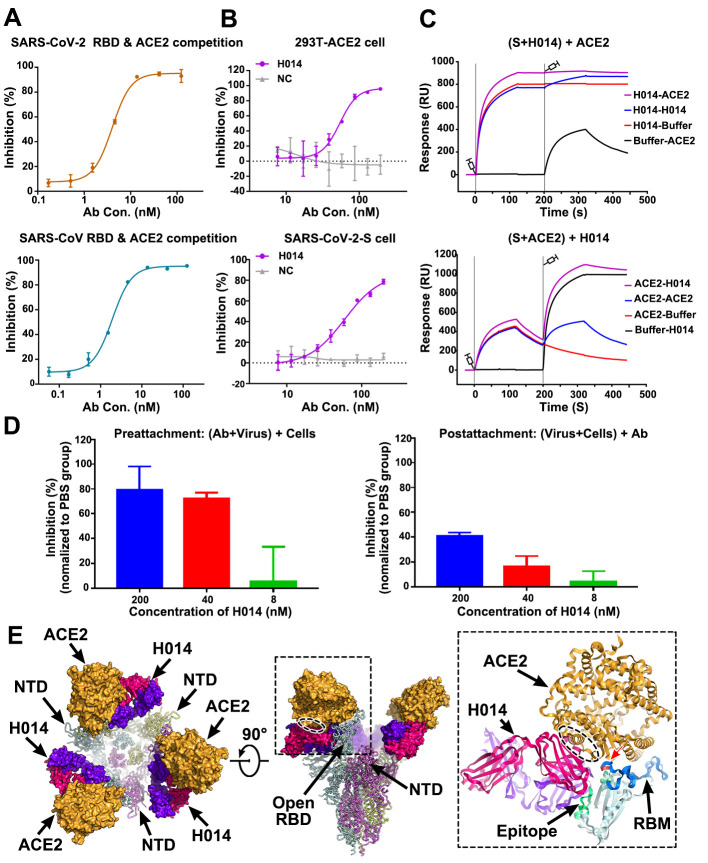
Fig. 3. Mechanism of neutralization of H014.
(A) Competitive binding assays by ELISA. Recombinant SARS-CoV-2 (upper) or SARS-CoV (lower) RBD protein was coated on 96-well plates, recombinant ACE2 and serial dilutions of H014 were then added for competitive binding to SARS-CoV-2 or SARS-CoV RBD. Values are mean ± SD. Experiments were performed in triplicate. (B) Blocking of SARS-CoV-2 RBD binding to 293T-ACE2 cells by H014 (upper). Recombinant SARS-CoV-2 RBD protein and serially diluted H014 were incubated with ACE2 expressing 293T cells (293T-ACE2) and tested for binding of H014 to 293T-ACE2 cells. Competitive binding of H014 and ACE2 to SARS-CoV-2-S cells (lower). Recombinant ACE2 and serially diluted H014 were incubated with 293T cells expressing SARS-CoV-2 Spike protein (SARS-CoV-2-S) and tested for binding of H014 to SARS-CoV-2-S cells. BSA was used as a negative control (NC). Values are mean ± SD. Experiments were performed in triplicate. (C) BIAcore SPR kinetics of competitive binding of H014 and ACE2 to SARS-CoV-2 S trimer. For both panels, SARS-CoV-2 S trimer was loaded onto the sensor. In the upper panel, H014 was first injected, followed by ACE2, whereas in the lower panel, ACE2 was injected first and then H014. The control groups are depicted by black curves. (D) Amount of virus on the cell surface, as detected by RT-PCR. Pre-attachment mode: incubate SARS-CoV-2 and H014 first, then add the mixture into cells (left); post-attachment mode: incubate SARS-CoV-2 and cells first, then add H014 into virus-cell mixtures (right). High concentrations of H014 prevent attachment of SARS-CoV-2 to the cell surface when SARS-CoV-2 was exposed to H014 before cell attachment. Values represent mean ± SD. Experiments were performed in duplicates. (E) Clashes between H014 Fab and ACE2 upon binding to SARS-CoV-2 S. H014 and ACE2 are represented as surface; SARS-CoV-2 S trimer is shown as ribbon. Inset is a zoomed-in view of the interactions of the RBD, H014 and ACE2 and the clashed region (oval ellipse) between H014 and ACE2. The H014 Fab light and heavy chains, ACE2, and RBD are presented as cartoons. The epitope, RBM and the overlapped binding region of ACE2 and H014 on RBD are highlighted in green, blue and red, respectively.

