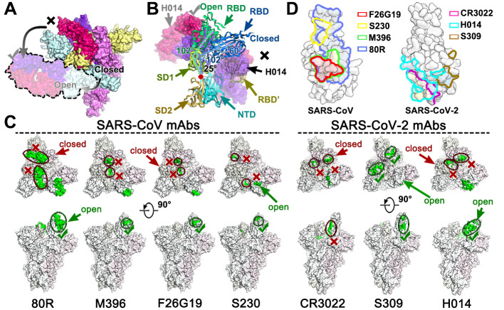
Fig. 4. Breathing of the S1 subunit and epitopes of neutralizing antibodies.
(A) H014 can only interact with the “open” RBD, whereas the “closed” RBD is inaccessible to H014. The “open” RBD and RBD bound H014 are depicted in lighter colors corresponding to the protein chain they belong to. Color scheme is the same as in Fig. 2A. (B) Structural rearrangements of the S1 subunit of SARS-CoV-2 transition from the closed state to the open state. SD1: subdomain 1, SD2: subdomain 2, RBD’: RBD (closed state) from adjacent monomer. SD1, SD2, NTD, RBD and RBD’ are colored in pale green, light orange, cyan, blue and yellow, respectively. The red dot indicates the hinge point. The angles between the RBD and SD1 are labeled. (C) Epitope location analysis of neutralizing antibodies on SARS-CoV and SARS-CoV-2 S trimers. The S trimer structures with one RBD open and two RBD closed from SARS-CoV and SARS-CoV-2 were used to show individual epitope information, which is highlighted in green. The accessible and in-accessible states are encircled and marked by green ticks and red crosses. (D) Footprints of the seven mAbs on RBDs of SARS-CoV (left) and SARS-CoV-2 (right). RBDs are rendered as molecular surfaces in light blue. Footprints of different mAbs are highlighted in different colors as labeled in the graph. Note: epitopes recognized by indicated antibodies are labeled in blue (non-overlapped), yellow (overlapped once) and red (overlapped twice).

