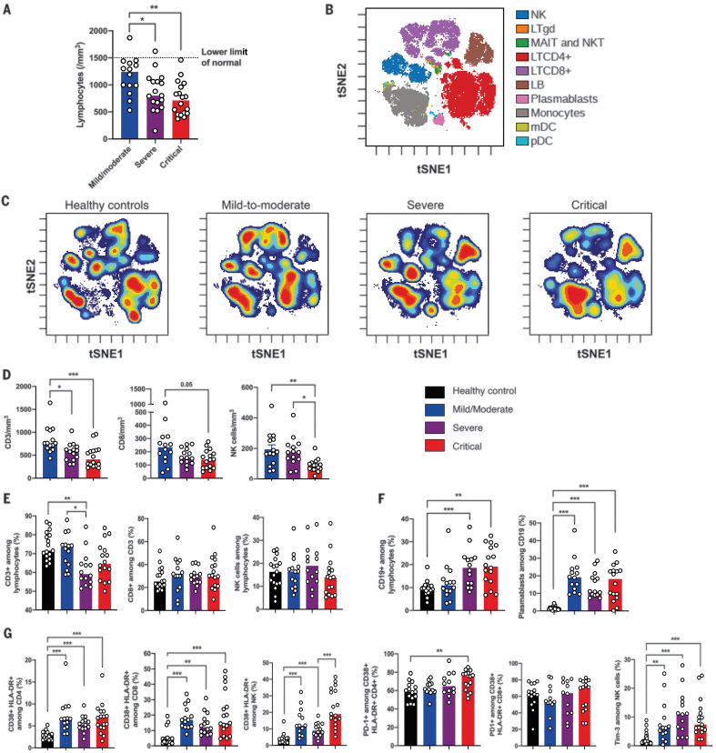
Fig. 1. Phenotyping of peripheral blood leukocytes in patients with SARS-CoV-2 infection.
(A) Lymphocyte counts in whole blood from COVID-19 patients were analyzed between days 8 and 12 after onset of first symptoms, according to disease severity. (B) viSNE map of blood leukocytes after exclusion of granulocytes, stained with 30 markers and measured with mass cytometry. Cells are automatically separated into spatially distinct subsets according to the combination of markers that they express. LTgd, γδ T cell; MAIT, mucosal-associated invariant T cell; LB, B lymphocyte. (C) viSNE map colored according to cell density across disease severity (classified as healthy controls, mild to moderate, severe, and critical). Red indicates the highest density of cells. (D) Absolute number of CD3+ T cells, CD8+ T cells, and CD3–CD56+ NK cells in peripheral blood from COVID-19 patients, according to disease severity. (E and F) Proportions (frequencies) of lymphocyte subsets from COVID-19 patients. (E) Proportions of CD3+ T cells among lymphocytes, CD8+ T cells among CD3+ T cells, and NK cells among lymphocytes. (F) Proportions of CD19+ B cells among lymphocytes and CD38hi CD27hi plasmablasts among CD19+ B cells. (G) Analysis of the functional status of specific T cell subsets and NK cells based on the expression of activation (CD38, HLA-DR) and exhaustion (PD-1, Tim-3) markers. In (D) to (G), data indicate median. Each dot represents a single patient. P values were determined with the Kruskal-Wallis test, followed by Dunn’s post-test for multiple group comparisons with median reported; *P < 0.05; **P < 0.01; ***P < 0.001.

