
Figure 1.
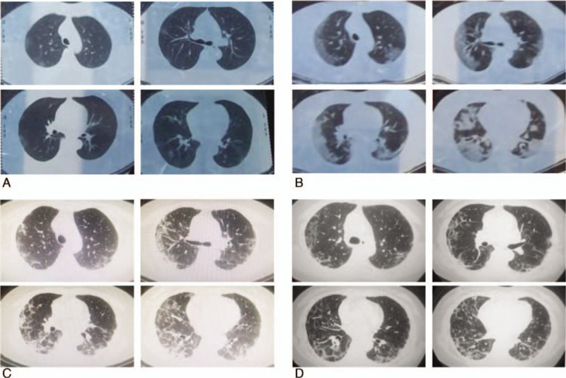
Serial chest computed tomography images over the course of the illness. A, Day 2: Ground-glass opacities are scattered peripherally in both lungs. B, Day 8: There is diffuse bilateral consolidation of the ground-glass opacities in both lungs. C, Day 18: The computed tomography (CT) image reveals partial resolution of the lung consolidation observed in the previous CT scan on Day 8. D, Day 23: The CT scan reveals almost complete resolution of the lung consolidation.