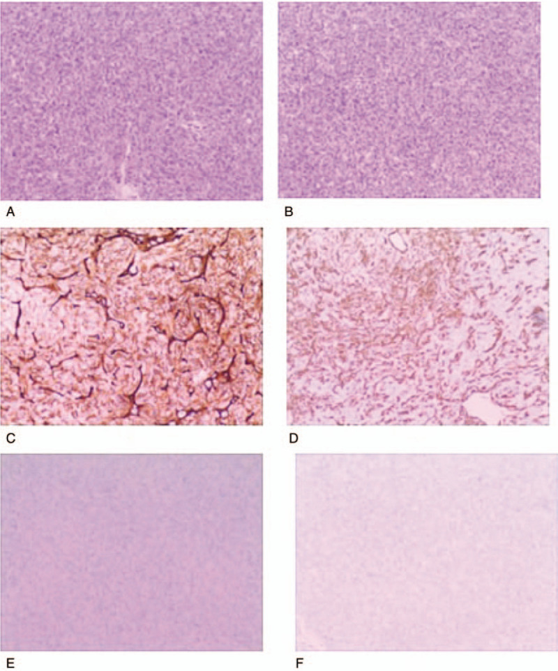
Figure 3.

Cells are arranged around thin-walled vascular spaces, with a staghorn appearance, [hematoxylin and eosin (H and E stain, original magnification ×200] (A and B), confirming the hypervascularity and non-epithelial origin of the neoplasms, which exhibited a mild nuclear pleomorphism, low mitotic rate (<5 mitoses/10 HPF), and few areas of necrosis. Immunohistochemical analysis was positive for CD34 (C) and vimentin (D), but negative for epithelial membrane antigen (E) and glial fibrillary acidic protein (GFAP) (F). H&E = hematoxylin and eosin.
