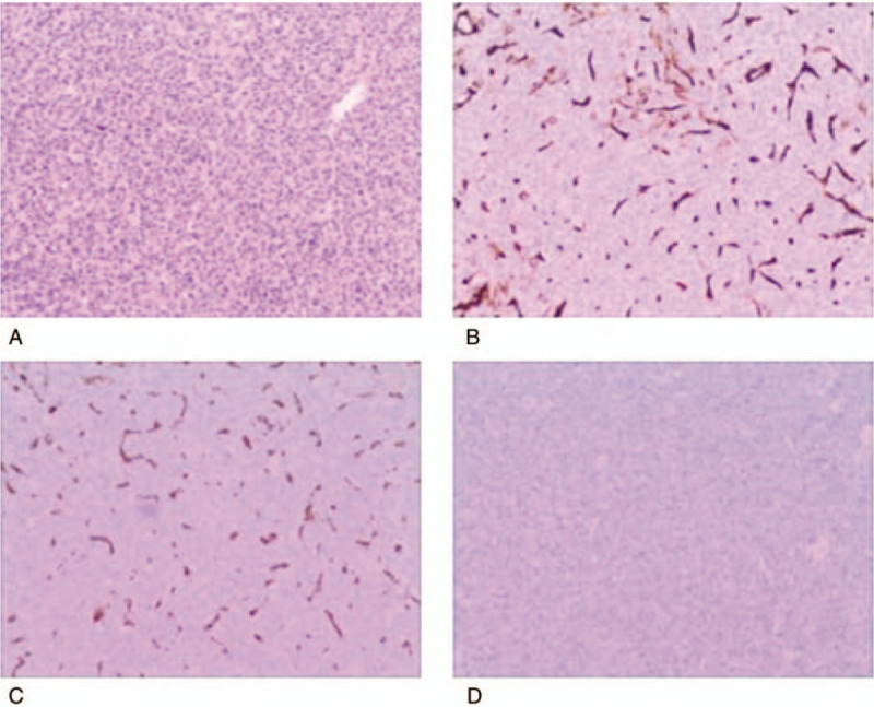
Figure 4.

Photomicrographs of the recurrent tumour in the bilateral frontal region. (A) The tumour consisted of cells with oval nuclei arranged in a turbulent pattern surrounding slit-like ‘staghorn’ vascular channels (H and E stain, original magnification ×200.) (B) Immunohistochemical examination revealing positive staining for D34 via sporadic patterns in tumour cells. Immunohistochemical analysis was partly positive for vimentin (C) and negative for glial fibrillary acidic protein (GFAP) (D). H and E = hematoxylin and eosin.
