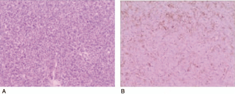
Figure 5.

(A) Photomicrographs of the recurrent brain tumour in the frontal region at 6 years before. Densely packed elongated or polygonal neoplastic cells are arranged around branching staghorn-like vasculatures (H and E staining) from the brain original tumour 6 years ago. (B) Ki-67 mitotic index showing a median value of 10%. H and E = hematoxylin and eosin.
