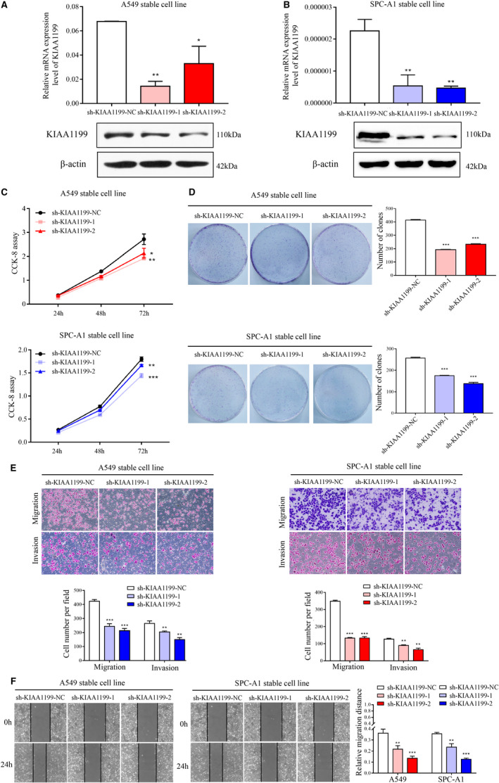
Figure 2.

Inhibition of NSCLC cell pathogenesis by KIAA1199 knockdown. A, and B, KIAA1199 mRNA and protein levels in KIAA1199‐knockdown cell lines. C, The cell proliferation of KIAA1199‐knockdown cells was assessed by CCK‐8 assay. D, The characteristic images of the cell colony formation were captured. The colonies were quantified in the graph on the right. E, The migration and invasion abilities were inhibited in KIAA1199‐knockdown cells. F, Wound closure was delayed in KIAA1199‐silenced cells compared with control cells in the wound healing assay. Each experiment was performed in triplicate. Significant differences: *P < .05, **P < .01, ***P < .001
