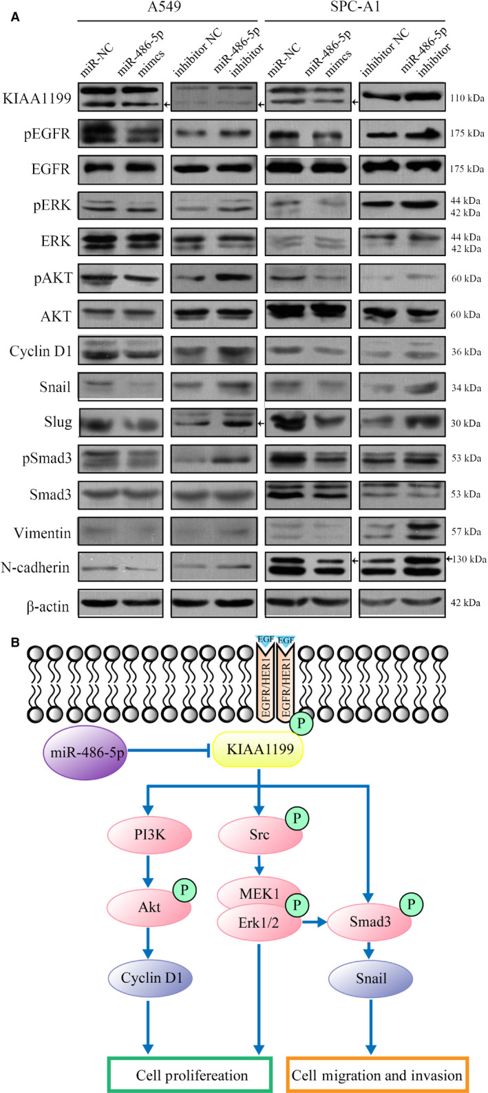
Figure 8.

A, A549 and SPC‐A1 cells were transfected with miR‐486‐5p mimics or inhibitor for 72 hours. The levels of pEGFR, EGFR, pErk, Erk, pAKT, AKT, pSmad3, Smad3, and other EMT markers were analyzed by western blotting. B, A diagram shows the mechanism of which miR‐485‐5p interacts with KIAA1199 and their control of EGFR‐related signaling pathways
