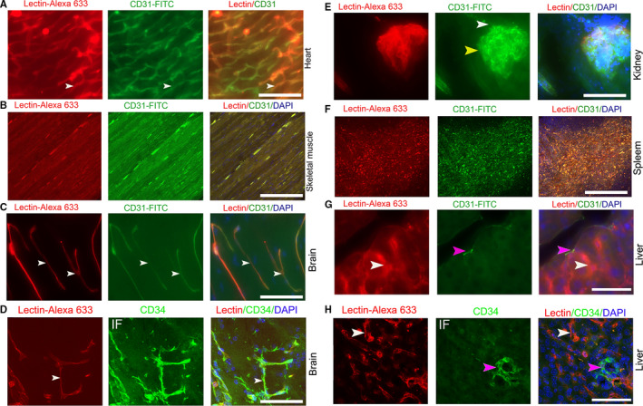
FIGURE 1.

The perfused staining of Lectin‐Alexa 633 and CD31‐FITC in normal murine organs. (A, B, C) Images of coperfused staining with CD31‐FITC and Lectin‐Alexa 633 in the heart, skeletal muscles, and brain of B6/C57 mice (white arrows, typical slender microvessels). (D) Comparison of coperfused staining of Lectin‐Alexa 633 in the brain with immunostaining with a CD34 antibody (white arrows, typical slender microvessels). (E) Images of coperfused staining with CD31‐FITC and Lectin‐Alexa 633 in the kidney (yellow arrow, glomerulus; white arrow, afferent artery). (F, G) Images of coperfused staining with CD31‐FITC and Lectin‐Alexa 633 in spleen and liver (pink arrow, central vein; white arrow, sinusoid endothelial cell). (H) Comparison of coperfused staining of lectin with CD34 antibody immunostaining in livers (white arrow, sinusoid endothelial). Mice, n = 3. IF, immunofluorescent staining. Scale bar, 50 µm
