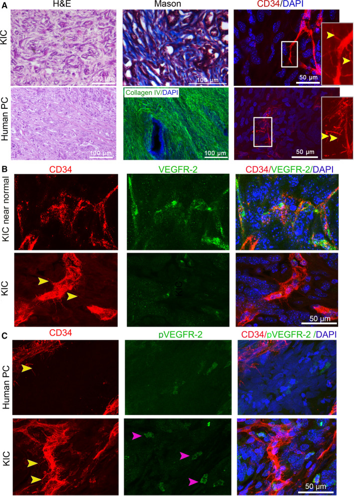
FIGURE 2.

Basal microvilli microvasculature in autochthonous PC in KIC resembles human basal microvilli microvasculature. (A) Comparison of the histology, stroma, and basal microvilli microvessels of the PC in KIC mice with human PC (yellow arrows, basal microvilli). A collagen IV antibody (green) stains human stroma. The inner panel, magnified region. (B) Comparison of VEGFR2 expression patterns in the basal microvilli microvessels of the PC in KIC with that in the near‐normal pancreatic tissue of KIC mouse (yellow arrows, basal microvilli). (C) Comparison of phospho‐VEGFR2Y996 (pVEGFR2Y996) expression levels in the basal microvilli microvessels of the PC in KIC with that in human PC (yellow arrows, basal microvilli). Mice, n = 2
