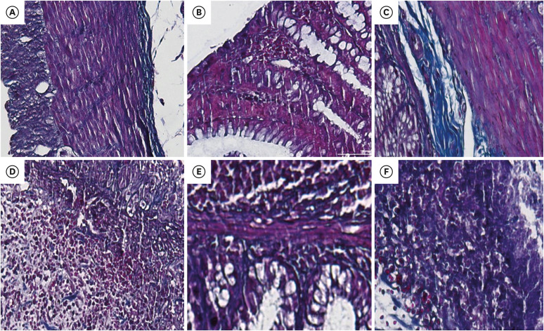
Figure 8. Photomicrographs of the rat colon stained Masson's trichrome stain (×200). The collagen deposition, showed by blue color staining area. Photomicrographs of protective group (A) AV 50 mg/kg, (B) AV 300 mg/kg, and treatment groups (C) AV 50 mg/kg, (D) AV 300 mg/kg, (E) positive control, (F) control group.
AV, Aloe vera.

