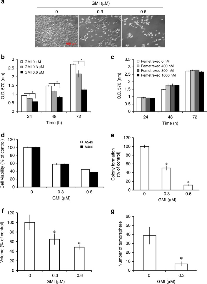
Fig. 1. Effects of GMI and pemetrexed on cell viability in A549/A400 pemetrexed-resistant lung cancer cells.
a After GMI (0, 0.3, and 0.6 μM) treatment for 48 h, the morphology of A549/A400 cells was investigated under an inverted microscope. Scale bar indicates 100 μm. b A549/A400 cells (2 × 10³ cells/well of 96-well plate) were treated with various concentrations of GMI (0, 0.3, and 0.6 μM) for 24, 48, and 72 h. Cell viability was analysed by MTT assay. c After treatment of pemetrexed (0, 400, 800, and 1600 nM) for an indicated time, MTT assay was performed to investigate the cell viability. d A549 and A549/A400 cells (2 × 10³ cells/well of 96-well plate) were treated with GMI (0, 0.3, and 0.6 μM) for 48 h. Cell viability was analysed by MTT assay. e A549/A400 cells (2 × 10² cells/well of six-well dish) were treated with various concentrations of GMI (0, 0.3, and 0.6 μM). After treatment for 24 h, the original medium was replaced with fresh medium, and the cells were incubated for 14 days for colony development. The number of colonies was counted under a dissecting microscope. The number of cells in each colony had to be larger than 50. Data show the relative colony number, and the number of cells without treatment was set at 100%. f A549/A400 cells (1 × 10³ cells/well of 96-well dish) were seeded onto ultra-low attachment 96-well plates. After 96 h incubation for spheroid formation, GMI (0, 0.3, and 0.6 μM)-containing medium was added to the well, and the spheroids were incubated for 7 days. Spheroids were investigated under an inverted microscope. The volumes of spheroids were determined by the formula 0.5 × larger diameter × small diameter2. Data show the relative spheroid volume, and the volume of spheroid without treatment was set at 100%. g A549/A400 cells (5 × 10³ cells/well of a six-well plate) cultured in the sphere formation medium with or without 0.3 μM GMI. After 14 days, the spheres were investigated and counted under an inverted microscope. The symbol ‘*’ indicates P < 0.05.

