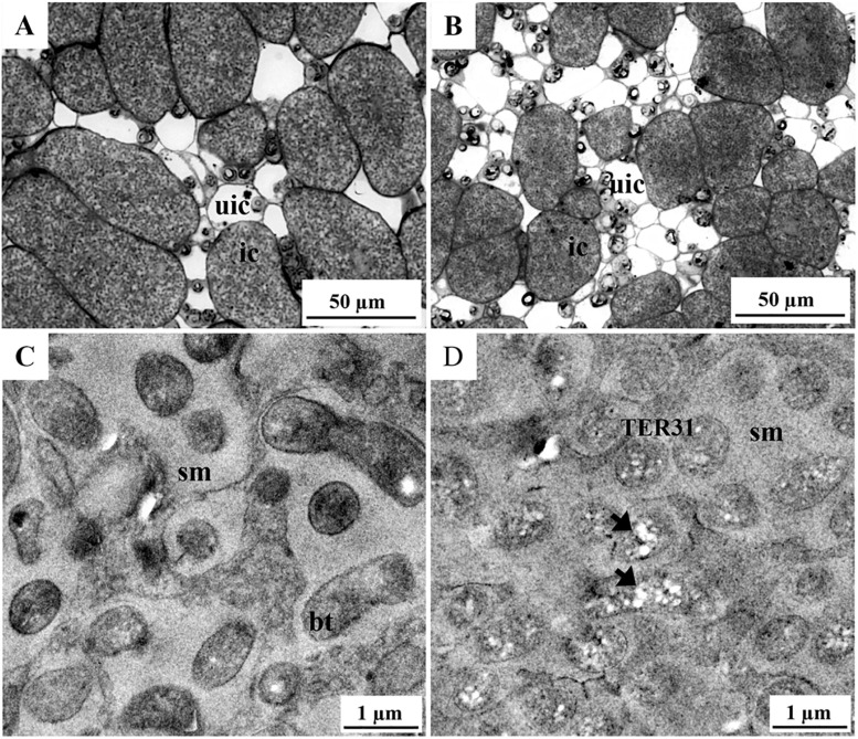
FIGURE 4.
Histology and transmission electron microscopy of infected cells from nodules generated on roots inoculated with TER31 and CFNX182-1 strains. Semi-thin and ultra-thin sections of nodule central tissue (21 dpi) were analyzed by bright field (A,B) and transmission electron microscopy (C,D). Samples: (A,C) nodules from roots inoculated with CFNX182-1 and (B,D) TER31. bt, bacteroids; ic, infected cells; sm, symbiosome matrix; TER31, intracellular TER31 bacteria; uic, uninfected cells; arrowheads, PHB-like storage material.

