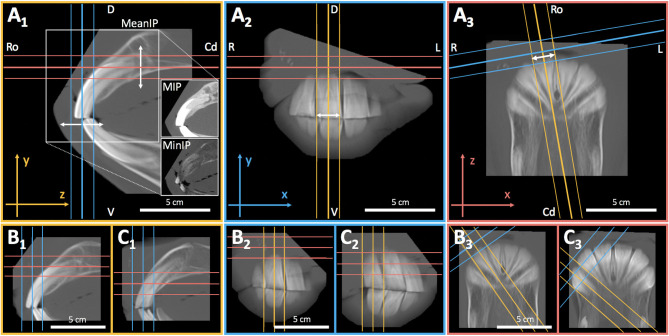
Figure 3.
3D multiplanar thick slab reconstruction (TSR) of individual incisors. TSR was used to merge contiguous slices within a certain range of the scan volume, hence creating a 2D x-ray-like sagittal image of each tooth. Superpositions with adjacent dental tissue structures could thus be reduced to a minimum. (A1) For optimal quality of TSRs the digital image calculation mode was set to “mean intensity projection” (MeanIP). Compare to the modalities maximum- (MIP) and minimum intensity projection (MinIP). (A–C2−3) Thick slab width was chosen according to the maximal mesiodistal tooth width of each single incisor (arrows) which was determined by running through the transverse and coronal plane of the tooth [arrows in (A1)]. The GRF allowed translational axis movements in the sagittal and transversal view (A–C1−2) as well as translational and rotatory axis movements in the coronal view (A–C3). The axes have been adjusted to reconstruct the tooth in its entire sagittal apicoocclusal extend without axial distortion (A–C1). Repeating this for each upper jaw incisor (B = 102, C = 103) and opposing lower jaw incisors resulted in 12 sagittal DICOM images per horse. R, right; L, left; D, dorsal; V, ventral, Ro, rostral; Cd, caudal.

