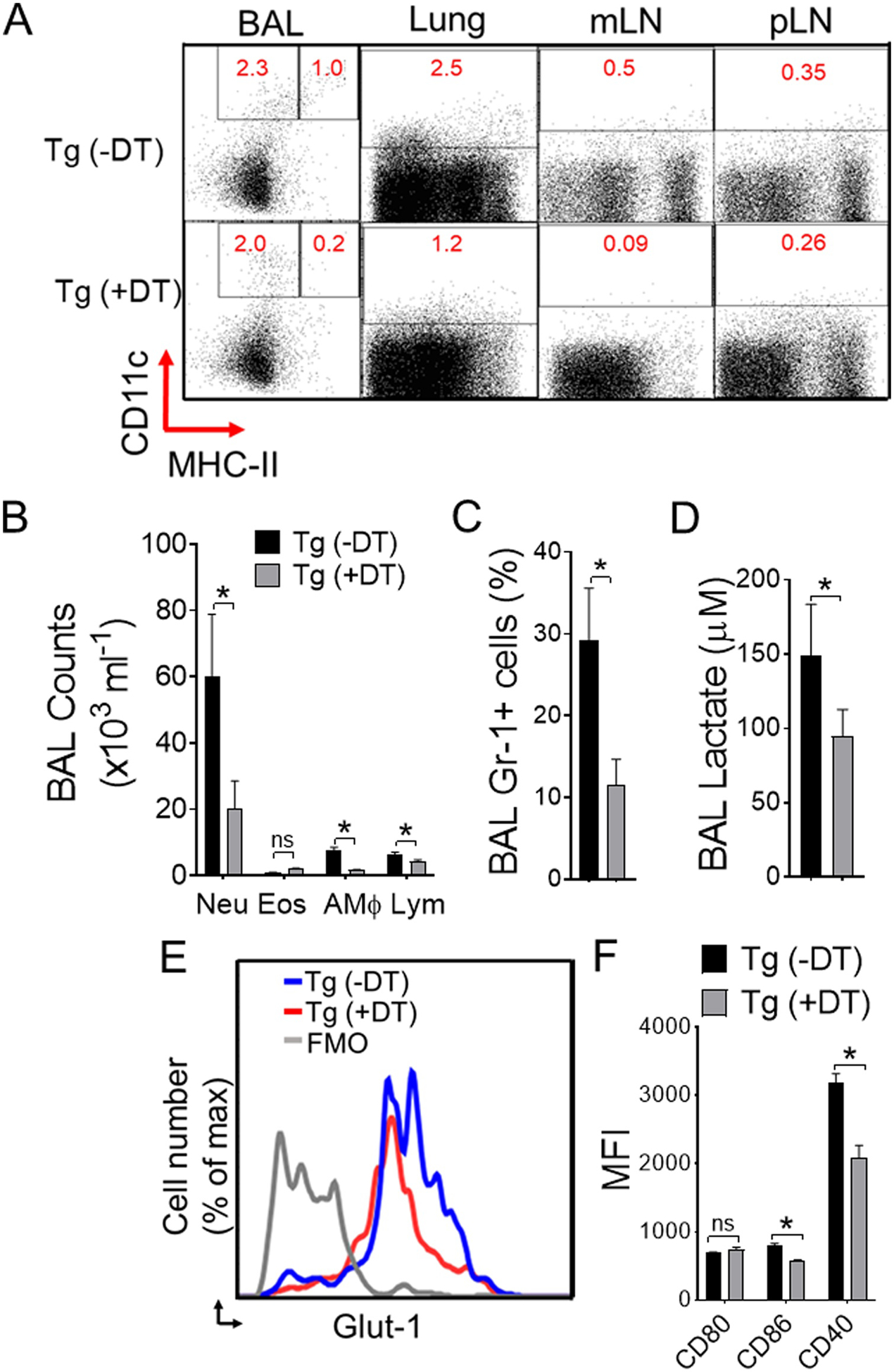
Figure 3. Lung CD11c+ APCs are critical regulator of Alternaria-induced airway inflammation.

(A) CD11c+ in BAL cells, lungs, lung draining mediastinal lymph node (mLN), and peripheral axillary lymph node (pLN) were analyzed by flow cytometry. Naïve CD11c-DTR transgenic (Tg) mice were injected or left untreated (PBS control) with DT (diphtheria toxin; 50 ng, i.n.) 24-hours before Alternaria challenge and analyzed at 6 hours. (B) Differential cell counts and (C) percentages of Gr-1+ neutrophils in BAL as measured by flow cytometry. (D) BAL lactate and (E) representative flow image of Glut-1 expression. Bar graph of (F) mean fluorescence intensity (MFI) of activation markers gated on lung CD11c+ CD11b+APCs from DTR mice ± DT treatment and receiving Alternaria. Results are expressed as mean ± s.e.m. Two independent experiments; n = 4 per group. Student’s t test *, P< 0.05.
