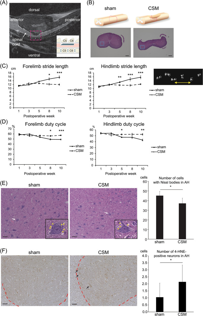
Figure 1.
Motor deficits and histopathologies of a rat model of CSM. (A) A representative T2-weighted mid-sagittal MRI image of a CSM rat at 10 weeks after surgery shows dorsal–ventral cervical spinal cord compression at C5 and C6 levels while keeping laminae of C4, 5, 6, and 7 intact. The area in the red dotted square is schematically shown in the inset. (B) Upper panels schematically show the spinal cord around the C5-6 level. Hematoxylin and eosin (HE) staining of axial sections at the red broken lines at the C5-6 disc level are shown in the lower panels. Blue boxed areas are enlarged in (E). Bar = 600 μm. (C,D) Stride length (C) and duty cycle (D) of fore- and hind-limbs analyzed by the CatWalk system of sham-operated and CSM rats from 1 to 10 weeks postoperatively. (C) A representative stride length of the right forelimb is indicated by a double-headed arrow in the right panel. (D) Duty cycle is a ratio in time when a paw stays on the glass plate in a single step cycle. CSM rats started to exhibit gait disturbance from 5 weeks after surgery. Mean and SE are indicated (n = 6 and 4 for sham-operated and CSM rats, respectively). *p < 0.05, **p < 0.01, and ***p < 0.001 by two-way repeated measures ANOVA followed by Bonferroni correction. (E,F) HE staining at 5 weeks postoperatively (E) and 4-hydroxynonenal (4-HNE) staining at 10 weeks postoperatively (F) of an axial section of the spinal cord at the C5-6 disc level in sham-operated and CSM rats. (E) Images are enlargement of the blue boxed areas in (B). A representative neuron (white arrow) in the anterior horn (AH) region is enlarged in the inset. Purple-stained Nissl bodies are indicated by yellow arrowheads. The numbers of cells with Nissl bodies in the AH region of sham-operated and CSM rats are shown. Mean and SD are indicated (n = 3 rats in each group). *p < 0.05 by Mann–Whitney U-test. (F) Representative images of the spinal cord stained with 4-hydroxynonenal (4-HNE). Arrows point to 4-HNE-positive large cells (diameter > 40 μm), which were recognized as motor neurons, in the AH region. A border between the gray and the white matters is indicated by a red dotted line. Bar = 50 μm. Mean and SD are indicated (n = 3 rats in each group). *p < 0.05 by Mann–Whitney U-test.

