Figure 3.
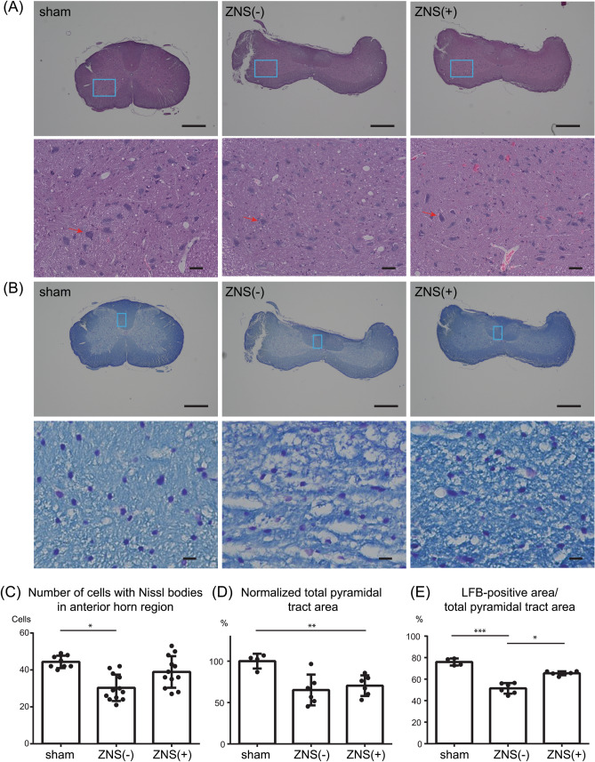
Effects of Zonisamide (ZNS) on the number of neurons in the anterior horn (AH) region and on demyelination of the pyramidal tracts. (A–B) Representative low (upper panels) and high (lower panels) magnification images of hematoxylin and eosin (HE) staining (A) and Luxol Fast Blue (LFB)/Nissl staining (B) of the spinal cord at the C5-6 disc level at 10 weeks after CSM operation. Blue squares in the AH region (A) and the pyramidal tract (B) in upper panels are magnified in lower panels. (A) A representative neuron in each panel is indicated by red arrow. Bars in upper panels = 600 μm. Bars in lower panels = 50 μm. (C) The number of cells with Nissl bodies in the AH region. (D) Total area of the pyramidal tracts was measured and normalized to that in sham-operated rat. The pyramidal tracts were compressed by a water-absorbing polyurethane blade in both ZNS(-) and ZNS( +) rats. (E) The area of LFB (light blue)-stained myelin was divided by the total area of pyramidal tracts in the spinal cord at the C5-6 disc level. (C,D,E) Mean and SD are indicated [n = 8, 12, and 12 anterior horns in 4 sham-operated, 6 ZNS(-), and 6 ZNS( +) rats, respectively]. * p < 0.05, ** p < 0.01, and *** p < 0.001 by Kruskal–Wallis test followed by Dunn-Bonferroni HSD.

