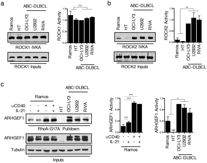
Figure 2.
Dysregulated activation of ROCK2 in ABC-DLBCLs. (a–b) ROCK1 and ROCK2 kinase activity was assayed by incubating immunoprecipitated ROCK1 (a) or ROCK2 (b) from nuclear extracts of BL, GCB-DLBCL, or ABC-DLBCL cell lines with purified recombinant MYPT1 as a substrate in the presence of ATP. Phosphorylated MYPT1 (pMYPT1) was detected using an antibody against pMYPT1. Total ROCK1 or ROCK2 input levels for each sample are shown in the lower panels. Quantifications are calculated as the densitometry ratio between pMYPT1 to total ROCK input protein (mean ± SEM; n = 2; p value by unpaired two-tailed t test). (c) RhoA-G17A-conjugated agarose beads were used to pull-down active ARHGEF1 from lysates of GCB-DLBCL, ABC-DLBCL, or Ramos cells following 6 h treatment with various combinations of αCD40 and IL-21. Quantifications are calculated as the densitometry ratio between ARHGEF1 from the RhoA-G17A pull-down to ARHGEF1 input levels [mean ± SEM; n = > 2; p value by 1-way ANOVA followed by Dunnett’s multiple comparisons test (left) or by unpaired two-tailed t test (right)]. * p < 0.05, ** p < 0.01, *** p < 0.001, **** p < 0.0001.

