. 2020 Jun 2;21(8):e48686. doi: 10.15252/embr.201948686

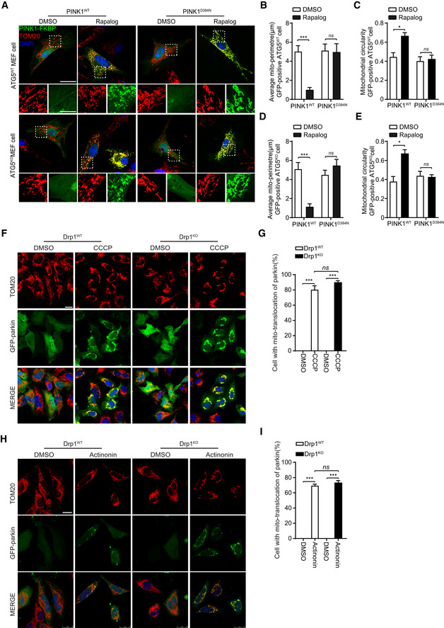
Figure 4. PINK1 phosphorylates Drp1S616 to regulate mitochondrial fission that is independent of mitophagy.

Figure 4

  • A–E
    ATG5 is dispensable for PINK1‐mediated mitochondrial fission. MEF cells derived from ATG5‐null (ATG5KO) and their wild‐type control mice (ATG5WT) were cotransfected Δ110‐PINK1‐GFP‐FKBP/FRB‐MTS with either PINK1WT or a kinase‐dead mutant PINK1D384N. Cells were induced with 250 nM rapalog (Rapalog) for 2 h to activate PINK1 kinase, followed by immunodetecting mitochondria (TOM20, red) and Δ110‐PINK1‐GFP‐FKBP/FRB‐MTS (PINK1‐FKBP, green). Cells treated with solvent (DMSO) were used as a treatment control. Representative images are shown, scale bar = 25 μm (A). Higher magnification images are also included (panels beneath the large cell images, scale bar = 10 μm). Mitochondrial morphology in different transfections was quantified (B–E). Student's test. *P < 0.05, ***P < 0.001, ns: no significance. Data were presented as mean ± SEM of three independent experiments. For each condition, > 100 cells were analyzed.
  • F–I
    Drp1 is dispensable for recruitment of parkin to mitochondria. Drp1WT and Drp1KO HeLa cells were transfected with GFP‐parkin. Cells were treated with either 20 μM CCCP for 2 h (F) or 150 μM actinonin for 6 h (H), followed by immunodetecting mitochondria (TOM20, red) and parkin (GFP‐parkin, green). Nuclei were labeled with DAPI (blue). Cells treated with solvent (DMSO) were used as a treatment control. Representative images were shown, scale bar = 25 μm. Cell with mitochondrial parkin (%) in different experimental group was quantified (G, I). One‐way ANOVA followed with Tukey's test. ***P < 0.001, ns: no significance. Data were presented as mean ± SEM of three independent experiments. For each condition, > 100 cells were analyzed.