
Figure EV1. Alpha‐lipoic acid (ALA) synthesis reduces in aged Drosophila midguts, and orally administered ALA rejuvenates aged intestinal stem cells (ISCs; related to Fig 1).
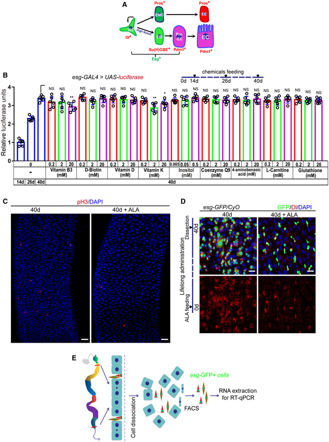
- Model of Drosophila intestinal stem cell (ISC) lineages. One ISC (Dl+ and Esg+) produces a new ISC and differentiates into a diploid precursor enteroblast (EB; Esg+ and Su(H)GBE+) with high Notch or a diploid precursor enteroendocrine mother cell (EMC). The EMC divides once to produce a pair of diploid enteroendocrine cells (EEs; Pros+). The post‐mitotic EB further differentiates into pre‐enterocyte (pre‐EC; Esg+ and Pdm1+), which continues to differentiate into an octoploid mature enterocyte (ECs; Pdm1+).
- Quantification of luciferase activity after administration of endogenous chemicals. Error bars show the SD of six independent experiments.
- Immunofluorescence images of pH3 staining with the midgut section from the R4 region in 40‐day flies and 40‐day flies with ALA administration started at 26th day after fly eclosion. pH3 (red) staining was used to visualize the mitosis of ISCs.
- Immunofluorescence images of esg‐GFP and Delta (Dl) staining with the midgut section from the R4 region in 40‐day flies and 40‐day flies with ALA administration after fly eclosion (lifelong administration). esg‐GFP (green) indicates ISCs and their differentiating cells. Dl (red) staining was used to visualize ISCs.
- A cartoon illustrating the sorting of esg‐GFP+ cells using FACS for RT–qPCR analysis (see Materials and Methods Details).