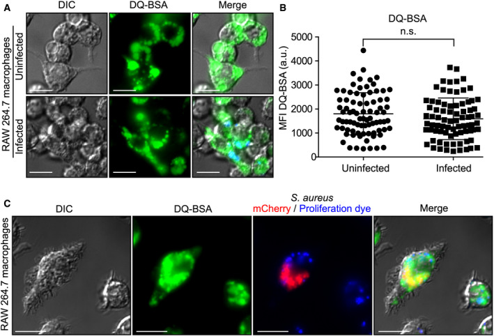
Figure 2. Infected macrophages can internalize and deliver degraded extracellular macromolecules to intracellular S. aureus .

- Internalization and processing of DQ™ Green BSA by uninfected and S. aureus (in blue)‐infected RAW 264.7 macrophages at 10 h post‐infection. These images are representative of three independent experiments.
- Quantitation of DQ‐BSA green fluorescence displayed by either uninfected or S. aureus (in blue)‐infected RAW 264.7. Shown is the mean fluorescence intensity (MFI) in arbitrary units (a.u) ± standard deviation where each symbol represents the measurement of a single cell. The data are derived from three independent experiments, and n.s. indicates the data are not significant as determined by an unpaired two‐tailed t‐test (P > 0.05).
- Fluorescence‐based proliferation assays were performed in conjunction with the probe DQ™ Green BSA. Processed DQ™ Green BSA could be found co‐localizing with intracellular mCherry‐expressing S. aureus (in red) and with intracellular S. aureus that has replicated (i.e., proliferation dye negative) at 10 h post‐infection as depicted here. Data information: Scale bars equal 10 μm.
