-
A, B
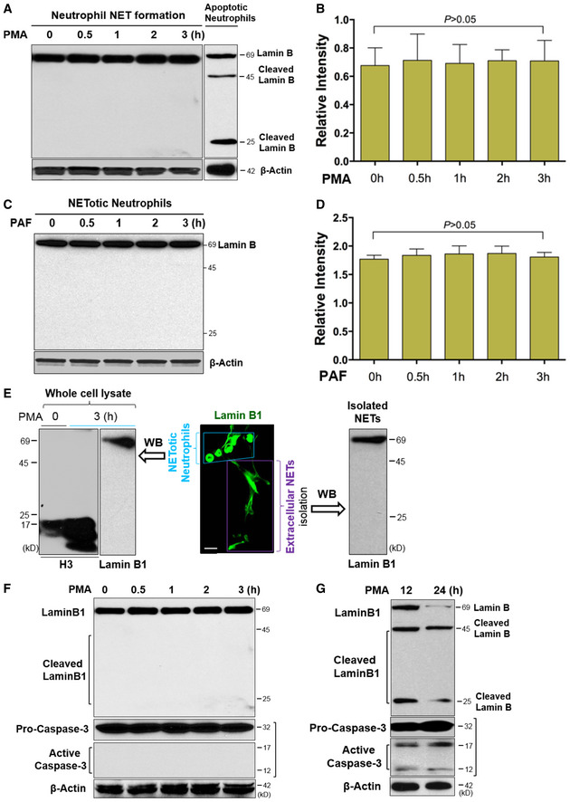
Representative and summary of immunoblots of full‐length lanes of lamin B in human dPMNs that were treated with PMA for 0, 0.5, 1, 2, 3 h during NET formation, or apoptotic dPMNs that were induced by PMA for longer term (12 h) treatment (A).
-
C, D
Representative and summary immunoblots of lamin B in human dPMNs that were treated with PAF for 0, 0.5, 1, 2, 3 h during NET formation.
-
E
Representative confocal microscopy image of a group of pPMNs with NET formation (enclosed by a light blue trapezoid) and their extracellular NETs (indicated by a purple rectangle square) released by these cells, as well as the immunoblot analysis of uncleaved lamin B and cleaved histone H3 with the whole‐cell lysates, as well as immunoblot analysis of uncleaved lamin B with lysate of the NETs isolated from conditioned medium of pPMNs with NET formation that were induced by 3 h PMA treatment.
-
F
Representative immunoblots with full‐length lanes for lamin B analysis, and pro‐caspase‐3 and its activated form caspase‐3 in human dPMNs with NET formation that were treated with PMA for 0, 0.5, 1, 2, 3 h.
-
G
Representative immunoblots display full‐length lamin B (69 kDa) and their cleaved fragments (45 and 25 kDa), and pro‐caspase‐3 and its activated form caspase‐3 in the apoptotic human dPMNs that were treated with PMA for 12, 24 h.
Data information: The anti‐human lamin B was used in all immunoblots (A–G) and the confocal image (E), and the latter was further detected by FITC‐labeled 2
nd Ab, scale bar, 20 μm. Anti‐human caspase‐3 was used (F, G), and β‐actin served as loading control (A–D, F, G). Data represent mean ± SD (
n = 3 biological replicates) for (B, D). Comparisons among three or more groups were performed using ANOVA, followed by Student–Newman–Keuls test.
Source data are available online for this figure.

