-
A–E
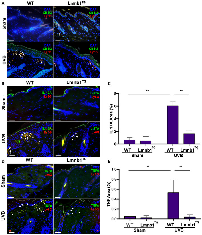
Fluorescent staining of the skin tissues from Lmnb1TG mice and their WT littermates that were irradiated or not (sham) by UVB. (A) The citrullinated histone H3 was probed by rabbit anti‐mouse citrullinated histone H3, following stained by Alexa Fluor‐488‐labeled donkey anti‐rabbit secondary antibody, while neutrophil marker was probed by rat anti‐mouse Ly6B Ab following stained by Alexa Fluor‐647 conjugated goat anti‐rat secondary antibody. (B–E) The representative (B, D) or summary (C, E) analysis of neutrophil surface marker Ly6G was stained by PE‐conjugated rat anti‐mouse Ly6G Ab. FITC‐labeled rat anti‐mouse IL‐17A antibody was used to detect IL‐17A. FITC‐labeled rat anti‐mouse TNF‐α antibody was used to detect TNF‐α. DNA was stained by DAPI for or panel (A–E).
Data information: White arrows indicate neutrophils with NET formation (A) or NETs with IL‐17A (B, C) or TNF‐α (D, E) display in skin. Scale bar, 100 μm. The panels (C, E) were calculated based on 3–4 individual mice (
n = 3–4 biological replicates). Data are given as mean ± SD. **
P < 0.01, between groups as indicated. Comparisons among three or more groups were analyzed using ANOVA, followed by Student–Newman–Keuls test.