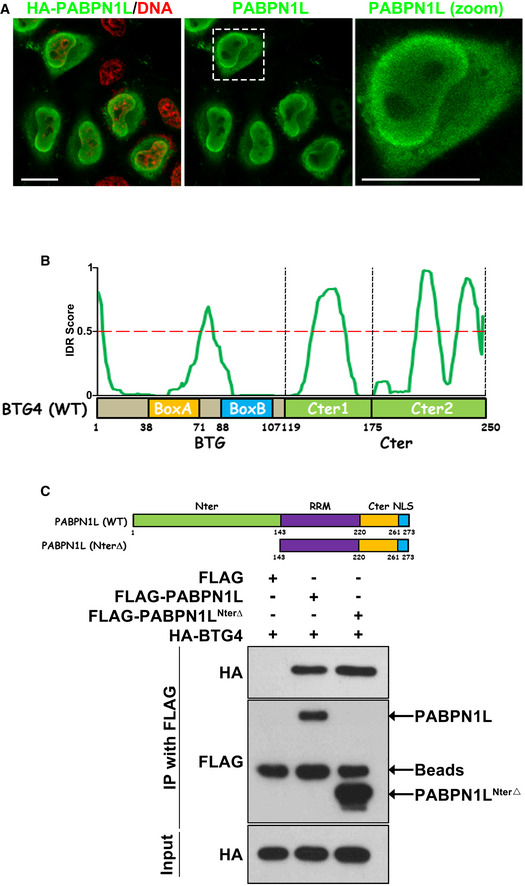
Figure EV3. PABPN1L interacts with BTG4.

-
AImmunofluorescent staining of HA (green) and DNA (red) in HeLa cells transfected with a HA‐PABPN1L expressing plasmid. The white dotted box showing the PABPN1L signal was zoomed out on the right panel. Scale bar = 10 μm.
-
BA diagram showing of mouse BTG4 with regions of predicted disorder.
-
CDiagrams and co‐IP results showing that BTG4 binds with PABPN1L and its N‐terminal‐deleted form (NterΔ). Lysates from HeLa cells expressing HA‐BTG4 and FLAG‐PABPN1L were immunoprecipitated with an anti‐FLAG antibody. At least three independent experiments were done with consistent results.
Source data are available online for this figure.
