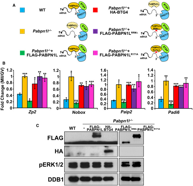
Figure 5. PABPN1L mediates BTG4 functions in vivo .

-
ADiagrams of the different treatments of (B).
-
BRT–qPCR results showing the relative expression level changes (MII/GV) of indicated transcripts. Fully grown GV oocytes of Pabpn1l −/− mice were microinjected with mRNAs encoding PABPN1L or BTG4 and were released from meiotic arrest at 12 h after microinjection. Total RNA was extracted from 10 oocytes in each sample. Error bars, SEM. *P < 0.05, **P < 0.01, and ***P < 0.001 by two‐tailed Student's t‐test compared to the first column. ns: non‐significant.
-
CWestern blot results showing the expression of exogenous PABPN1L (full length, RRM‐deleted, or R171A mutant) and BTG4 in Pabpn1l −/− oocytes at the MII stage. The total protein from 100 oocytes is loaded in each lane. pERK1/2 is blotted to indicate the developmental stages. DDB1 is blotted as a loading control. At least three independent experiments were done with consistent results.
Source data are available online for this figure.
