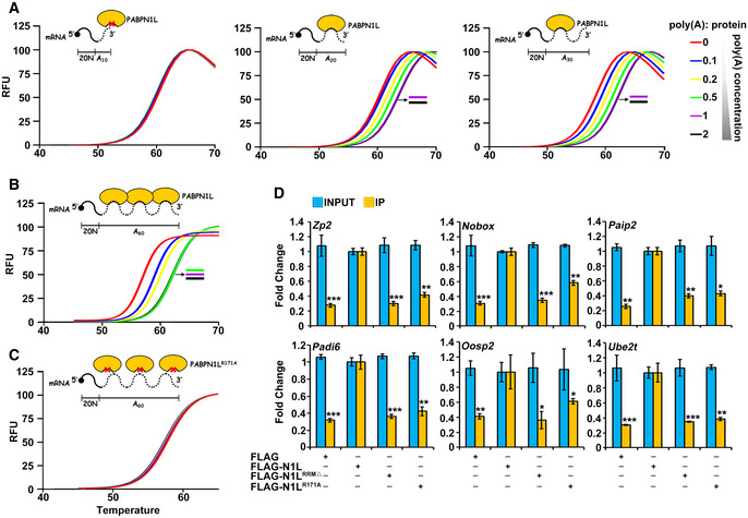
Figure 6. Characterization of the PABPN1L poly(A) binding ability.

-
A–CDiagrams and averaged fluorescence traces of the thermal stability assays of PABPN1L in the presence and absence of poly(A) variants (n = 6). The RNA substrate comprised 20 non‐poly(A) nucleotides followed by a 10/20/30/60‐adenosine poly(A) tail. PABPN1L‐bound substrates were prepared with 2/1/0.5/0.2/0.1 RNA molecules per PABPN1L. The midpoint of the unfolding curve was determined as the melting temperature (Tm).
-
DRNA immunoprecipitation results showing the interaction of PABPN1L with indicated transcripts. Fully grown GV oocytes of WT mice were microinjected with mRNAs encoding PABPN1L (WT, RRM‐deleted, or R171A mutant) and were released from meiotic arrest at 12 h after microinjection. Total RNAs were extracted from 350 MI stage oocytes in each sample. RNAs recovered from the immunoprecipitants were subjected to RT–qPCR of the indicated transcripts. Fold change values of both Input and IP samples were normalized by RIP results of the FLAG‐PABPN1L microinjection group. Error bars, SEM. *P < 0.05, **P < 0.01, and ***P < 0.001 by two‐tailed Student's t‐test comparing with RNA levels pulled down by PABPN1L WT in the second column.
