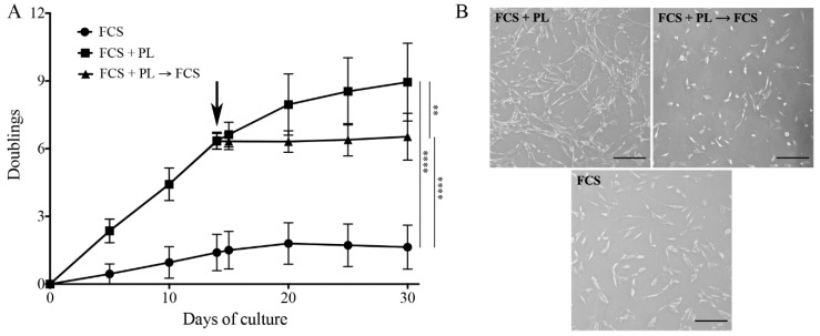
Figure 1.
Platelet lysate (PL) induces proliferation of “aged” osteoblasts. (A) Growth kinetics plotted as number of cell duplications versus time of culture. At T0, a culture of primary osteoblasts maintained in standard medium in the presence of fetal calf serum (FCS) for 5 passages was split in two and the cultures continued in the presence and in the absence of PL. At T14 (arrow) the PL culture was again split in two and the cultures continued in the presence and in the absence of PL. At each culture time, cells were detached and counted by a hemocytometer. Three independent experiments on 3 different primary cultures were performed. At each time, cell counting was done in duplicate dishes. The average values ± standard error of the mean (SEM) are shown. The significant differences in proliferation rate are also shown, ** p < 0.01 and **** p < 0.0001. (B) Representative images of osteoblasts in the different culture conditions. Scale bar = 50 µm.

