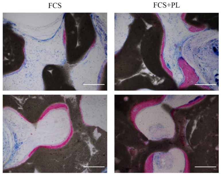
Figure 3.
Osteogenic differentiation potential of PL treated and untreated osteoblasts in vivo. Histological analysis by Stevenel’s/Van Gieson staining of ectopic tissue formed after subcutaneous implantation in nude mice of osteoblasts expanded in standard culture medium (left panels) or osteoblasts expanded standard culture medium supplemented with PL (right panels) seeded on osteoinductive scaffolds. 1.5 × 10⁶/scaffold (Upper panels) or 2.5 × 10⁶ cells/scaffolds (lower panels) were implanted. The purple stain refers to the newly deposited calcified bone and the pale pink the non, or only poorly, calcified osteoid (still immature bone). In blue non bone tissues. Scale bar = 200 µm.

