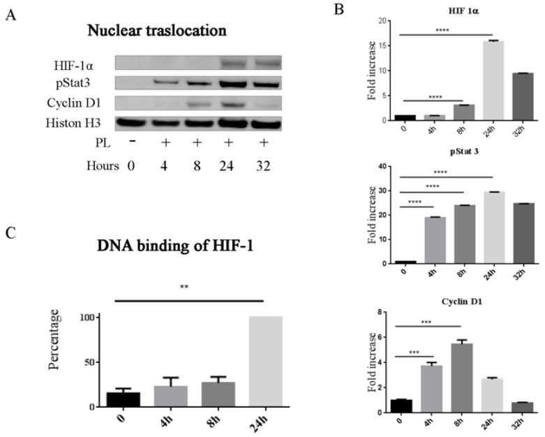
Figure 5.
PL promotes stabilization, activation and nuclear translocation of HIF-1α and pStat3. (A) Western blot analysis of nuclear proteins extracted from PL treated and control untreated cells. Cyclin D1 and Histone H3 were analyzed in parallel as controls. (B) Quantification of HIF-1α, pSTAT3, and Cyclin D1 present in cell nuclei. Density values are referred to histone H3 and shown as fold increase (n = 3); at 32 h, only two samples were analyzed. (C) Binding to hypoxia-response element (HRE) sequence of the active HIF-1α complexes in nuclear extracts. Data are shown as the percentage of average Optical Density (OD) value at 450 nm of the cells at different times with respect to treated cells at 24h (100%). Two independent experiments were performed in duplicate starting from two different primary cultures. ** p < 0.01, *** p < 0.001, and **** p < 0.0001.

