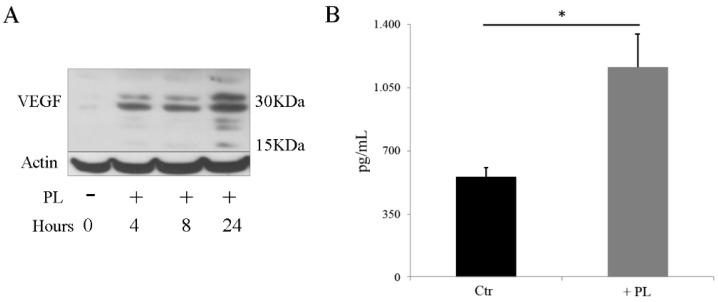
Figure 7.
PL induces the expression of vascular endothelial growth factor (VEGF). (A) Western blot analysis of proteins extracted from cells expanded in medium containing FCS and transferred in SF medium and in the presence of PL for different times. (B) Quantification of VEGF secreted in the cell culture medium. After cell expansion in medium containing FCS, the control culture (Ctr) was transferred and maintained in SF medium whereas the other culture was transferred in SF medium for 2 h and then moved to SF medium supplemented with 5% PL. After 24 h, both cultures were washed with PBS and further incubated with SF + 0.1% FCS for 16 h. Supernatants were collected and VEGF concentration was determined. Results are expressed as pg/mL of supernatant. Two independent experiments were performed on two different primary cultures. Determinations were performed in duplicate at two different dilutions for each experiment. * p < 0.05.

