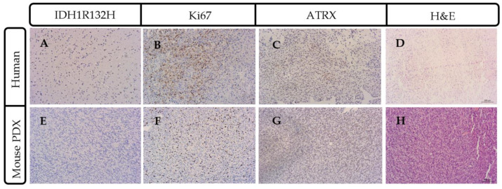
Figure 3.
Parental patient tumor mutational status was recapitulated in PDX mouse model. Representative case 2409 lacked IDH1R132H consistent with the wildtype IDH1 molecular characterization (A) that was also present in the mouse model of glioma (E). Ki67 staining indicated a highly-proliferative tumor in the parental (B) and mouse PDX (F) model. ATRX (ATP-dependent helicase, X-linked protein) immunohistochemistry had a similar pattern of expression in the human (C) and the mouse (G). (D) ATRX and hematoxylin and eosin (H&E) staining of parental human patient tumor and (H) dense nuclei with pseudo-palisading necrosis is present in the mouse intracranial PDX model. Scale bar = 100 μm.

