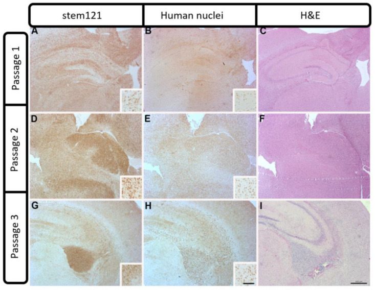
Figure 4.
Patient-derived PDX model of case 2409 showing diffuse STEM 121 staining of human-derived cells in passages 1 and 2 (A,D), and a more localized staining with some invasion in passage 3 (G). An adjacent section of human nuclei staining showed the same pattern of diffuse staining in passages 1 and 2 (B,E) and a localized staining with some invading cells evident in passage 3 (H). Increased cellularity and infiltration of proliferating tumor cells were apparent in H&E stained sections Insets (C,F,I) show individual cellular staining. Scale bar = 500 μm. Inset scale bar = 100 μm).

