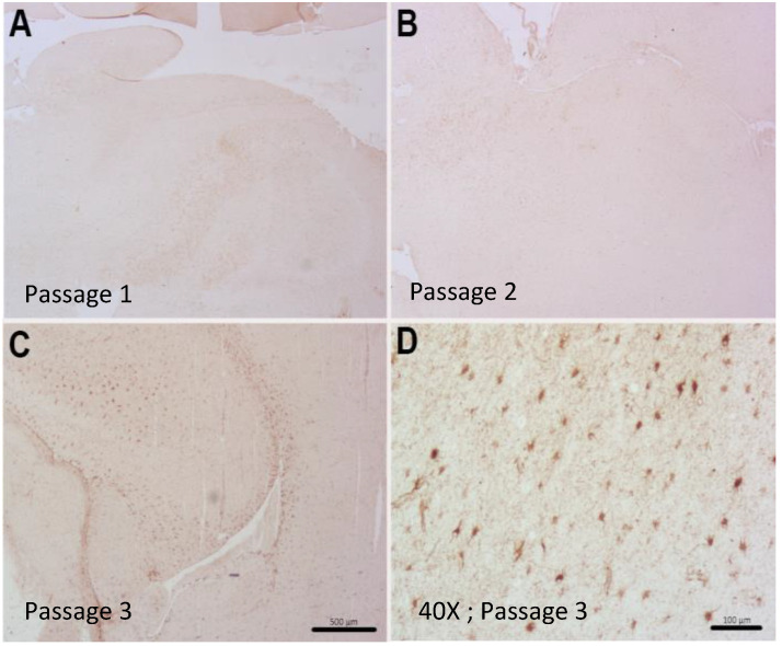
Figure 5.
Patient-derived intracranial PDX model of case 2409 showed reactive astrogliosis with glial fibrillary acidic protein (GFAP) staining in the third passage (C) and little to no glial staining in the first two passages (A,B). (D) Tumor sample from the patient demonstrating reactive gliosis. Scale bars = 500 μm (A–C) and 100 μm (D).

