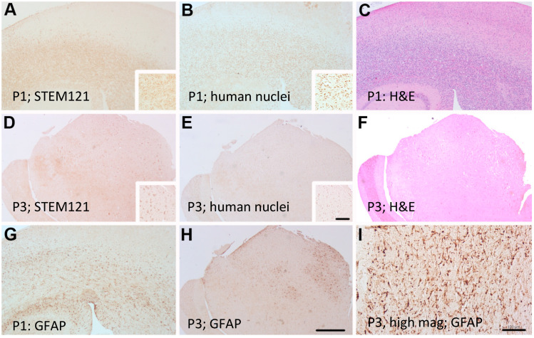
Figure 6.
Adjacent sections of patient derived PDX model of case 2025 showing diffuse STEM 121 staining in passages 1 (P1) and 3 (P3) (A,D) respectively and diffuse human nuclei staining in passage 1 (P1) and 3 (P3) (B,E) respectively. GFAP staining showed reactive astrogliosis (G,H). Resected human tumor sample from the patient illustrates reactive gliosis (I). H&E staining clearly shows the increase tumor cell proliferation (C,F). Insets show individual cellular staining. Scale bar = 500 μm (A–H). Scale bar = 100 μm (I) Inset scale bar = 100 μm.

