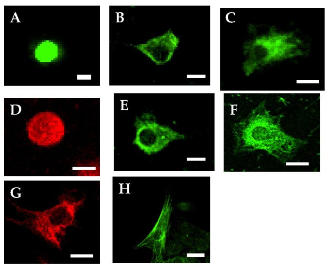
Figure 1.
Immunofluorescent images of naïve epidermal stem cells or spontaneously differentiated cells. (A), p75NTR stain; (B), fibronectin stain; (C), nestin stain; (D), ret proto-oncogene product (RET) stain; (E), Keratin stain; (F), glial fibrillary acidic protein (GFAP) stain; (G), Neurofilament-M stain; (H), Smooth muscle actin stain. Fluorescein isothiocyanate (FITC), green; TRIC, red; Scale bar = 10 μm.

