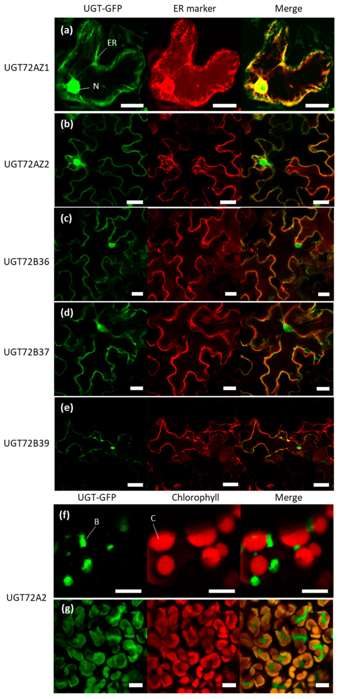
Figure 9.
Subcellular localization of poplar UGT72 fused to GFP. (a–f) UGT72-GFP fusion proteins and 35S::AtWAK2-mCherry, as ER marker (a–e) in N. benthamiana leaf epidermal cells after agroinfiltration. UGT72A2-GFP (f) was localized in bodies associated to chloroplasts (red fluorescence). (g) Stable transgenic poplar line expressing the same 35S::UGT72A2-GFP construct. UGT72A2 was localized in in bodies associated with chloroplasts and possibly in chloroplasts. Images are representative of biological triplicates. B, bodies associated with chloroplast; C, chloroplast; ER, endoplasmic reticulum; N, nucleus. Scale = 20 µm (a), 35 µm (b–d), 70 µm (e), 5 µm (f), 10 µm (g).

