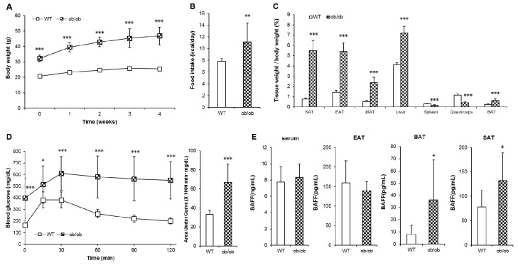
Figure 5.
ob/ob mice shows deteriorated glucose tolerance with tissue-specific increase of BAFF expression. Leptin-deficient (ob/ob) mice and C57BL/6J (wild-type, WT) mice were maintained on a normal chow diet for 5 weeks. (A) Changes of body weight over 5 weeks (n = 10). Changes of (B) average calorie intake and (C) tissue weights (n = 10). (D) Glucose tolerance test of WT and ob/ob mice (n = 10). Mice fasted for 16 h, and the blood glucose levels were measured at 0, 15, 30, 60, 90 and 120 min after intraperitoneal injection of glucose (2 g/kg). (E) Protein BAFF concentration in serum and adipose tissues (n = 8). Data represent means ± SD. * p < 0.05, ** p < 0.01, *** p < 0.001 between wild-type and ob/ob mice. SAT: subcutaneous adipose tissue, EAT: epididymal adipose tissue, MAT: mesenteric adipose tissue, BAT: interscapular brown adipose tissue.

