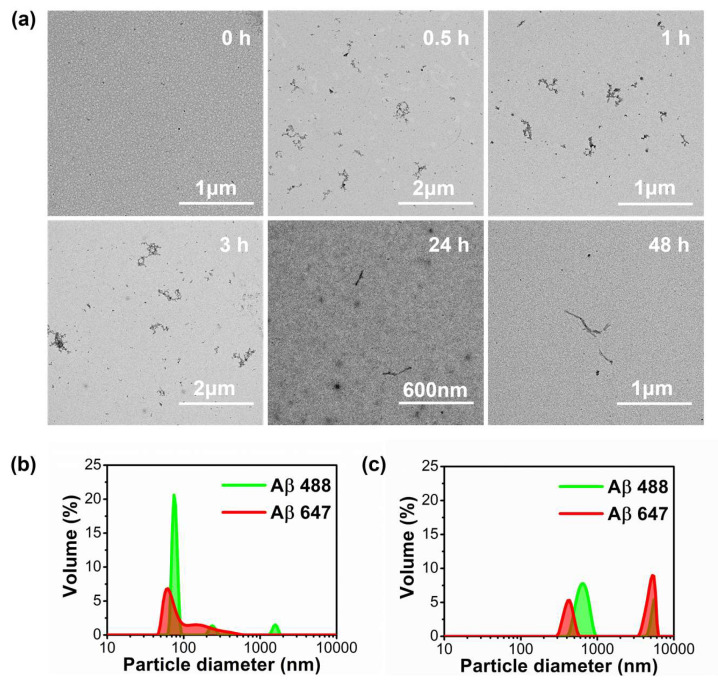
Figure 3.
(a) Transmission electron microscopy (TEM) images of fluorescently labeled Aβ aggregates and fibers (equimolar 0.5 μM mixture of Aβ-488 and Aβ-647) at different incubation times. (b,c) dynamic light scattering (DLS) images of labeled Aβ peptides (Aβ-488: green line and Aβ-647: red line) formed at 0 h (b) and 0.3 h (c) of the incubation process, sonicated in a quartz cuvette in an NH3 solution (pH 12) to avoid their aggregation (b) and in a phosphate buffered saline solution pH 7.4 after 20 min of aggregation (c).

