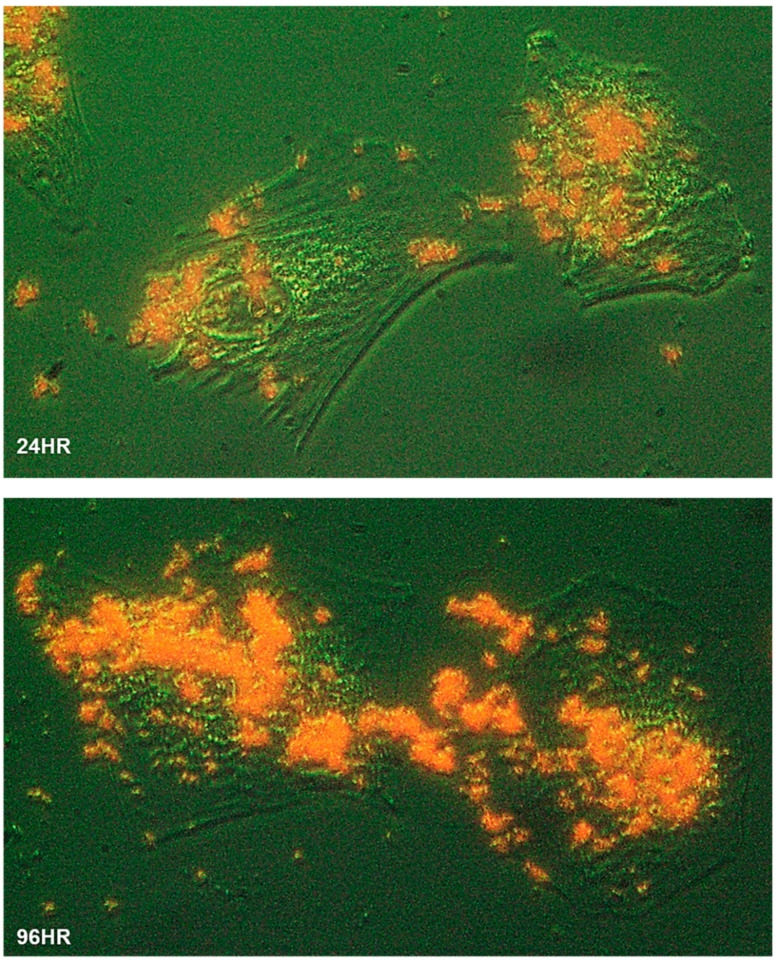
Figure 2.
Biomineralization of human cells. The Saos-2 cells grew at low confluency on a glass slide immersed in MEMα/10% FBS with αGP (2 mM) for 24 h (24HR) and 96 h (96HR), respectively. The slides were washed, fixed, and stained with Alizarin Red S. The cell morphology was illustrated using optical phase-contrast (with pseudo-green background), and HAP minerals were in red under a ZEISS inverted microscope equipped with an Infinity 3 digital camera and imaging software. Cells were magnified 400×.

