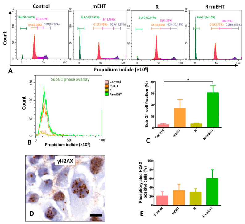
Figure 5.
Treatment related elevated subG1-fractions and moderate increase in nuclear phosphorylated H2AX protein levels in Panc1 cultures 24 h post-treatment. Representative flow cytometry graphs of cell cycle fractions (A). A summary (B) and graphical representation of subG1-phase fractions (C) showing strong significance among the 4 groups (p = 0.0006), which was confirmed with the Dunn’s test between R+ mEHT treatment and the controls (p = 0.028). Upregulated granular γH2AX foci in tumor cell nuclei after combined R+mEHT treatment (D, scale bar: 10 µm) and their graphical representation after quantification (E), whose difference, however, did not reach significance compared to the controls. The asterisk (*) marks the value of p ≤ 0.05.

