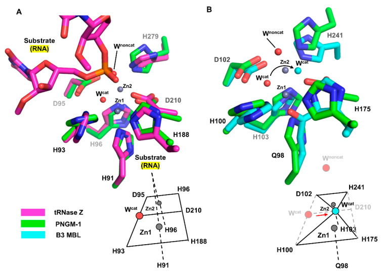
Figure 3.
Comparison of the MBSs between PNGM-1 and Bs-tRNase Z and B3 MBL GOB-18. (A) The MBS of PNGM-1 (green) superimposed on that of Bs-tRNase Z (magenta). The metal ions (grey spheres) and water molecules (red spheres) belong to the structure of wild-type PNGM-1. (B) The MBS of PNGM-1 (green) superimposed on that of B3 MBL GOB-18 (cyan). The metal ions (grey spheres) and red water molecules belong to the structure of wild-type PNGM-1, whereas the cyan water molecule belongs to that of GOB-18. Metal coordination schemes are shown at the bottom.

