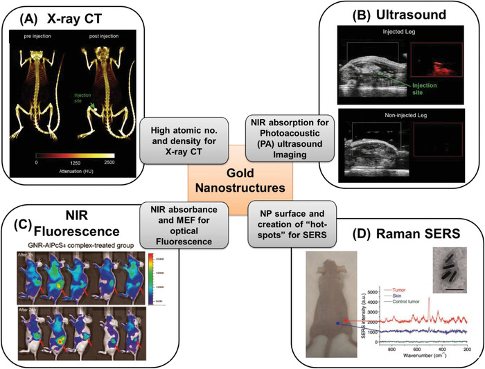
Figure 8.

Examples of gold nanostructure employed in optical diagnosis in mice tumor models by injecting the customized gold nanostructures, along with featuring their specific property for specific diagnosis. A) X‐ray CT imaging demonstrating tumor detection (marked with arrow) for pre‐ and postinjection. Adapted with permission.[ 106 ] Copyright 2016, Elsevier. B) Ultrasound imaging demonstrating tumor detection (marked with arrow) in injected and noninjected leg of mice. Adapted with permission.[ 106 ] C) NIR fluorescence imaging with coated gold NRs after 1 and 24 h demonstrating tumor detection (marked with arrow). Adapted with permission.[ 107 ] Copyright 2011, American Chemical Society. D) Raman (SERS‐based) spectroscopy employing gold NRs demonstrating tumor detection by comparing the spectrum of skin, tumor, and control tumor. Adapted with permission.[ 108 ] Copyright 2010, Wiley.
