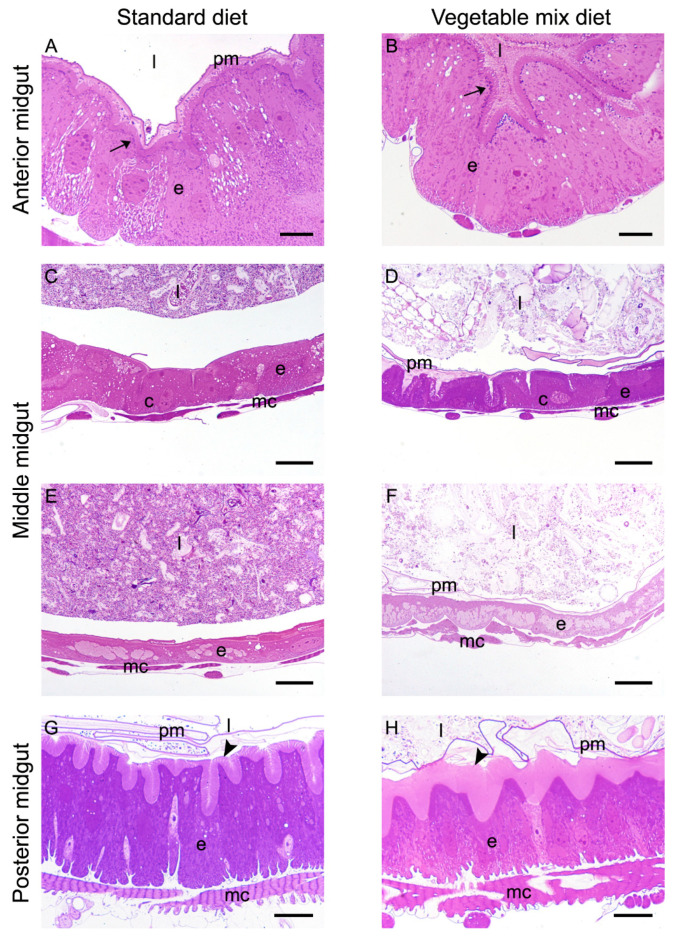
Figure 3.
Morphological comparison of midgut from larvae reared on SD and VMD. (A,B): cross-sections of the anterior midgut. (C–F): copper (C,D) and large flat cells (E,F) in the middle midgut of H. illucens larvae. (G,H): cross-sections of the posterior midgut. Columnar cells of larvae grown on VMD (H) show microvilli (arrowheads) that are longer than those of columnar cells of larvae reared on SD (G). Arrows: dark vesicles under the brush border. c: copper cells; e: epithelium; l: lumen; mc: muscle cells; pm: peritrophic matrix. Bars: 10 μm (A,B), 20 μm (C–H).

