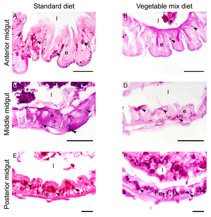
Figure 4.
Comparison of glycogen accumulation in the three midgut regions of larvae reared on SD and VMD—Periodic Acid-Schiff (PAS) staining. (A,B): anterior midgut of larvae reared on SD (A) shows a higher accumulation of glycogen (arrowheads) than VMD (B). (C–F): middle (C,D) and posterior (E,F) midgut of larvae reared on the two diets do not show significant differences in glycogen accumulation. e: epithelium; l: lumen. Bars: 50 μm (A,B,E,F), 20 μm (C,D).

