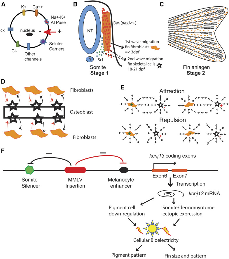
Figure 9.
Model of kcnj13 regulation in the Dhi2059 mutant and fin patterning by bioelectricity. (A) Illustration of cell bioelectric dynamic properties, which may be contributed by potassium channels (K+); calcium channels (Ca++), chloride channels (Cl−), solute carriers, connexins (cx), and Na+-K+ ATPases. (B–E) Two-stage bioelectric model of zebrafish fins. (B) The first stage of bioelectric regulation happens among the fin progenitor cells within a somite. The dashed lines indicate that the possible source of the second wave progenitors may come from the myotome or sclerotome. The bioelectric alteration of either the first-wave progenitor cells (orange dots) or the second-wave progenitor cells (orange or blue dots) cause fin patterning changes. (C) The second stage of bioelectric regulation happens within the fin anlagen. (D) The interaction of the fibroblasts and osteoblast eventually determine fin size and pattern. (E) Either attraction or repulsion occurs between fibroblasts and osteoblasts, depending on their dynamic bioelectric status. (F) A model of kcnj13 regulation by retroviral insertion in the Dhi2059 mutant. The Moloney murine leukemia virus is inserted within exon 5 of the kcnj13 allele in Dhi2059 fish. This viral insertion may negatively affect the melanocyte-specific enhancer and somite/dermomyotome-specific silencer, either through physical distance isolation or viral long terminal repeat regulation. Thus, this insertion leads to ectopic somite expression and slightly reduced expression in the melanocyte. This leads to cell bioelectricity change and subsequent patterning alterations in corresponding organs. DM, dermomyotome; dpf, days postfertilization; mRNA, messenger RNA; n, notochord; NT, Neural tube; Scl, sclerotome.

