Figure 6.
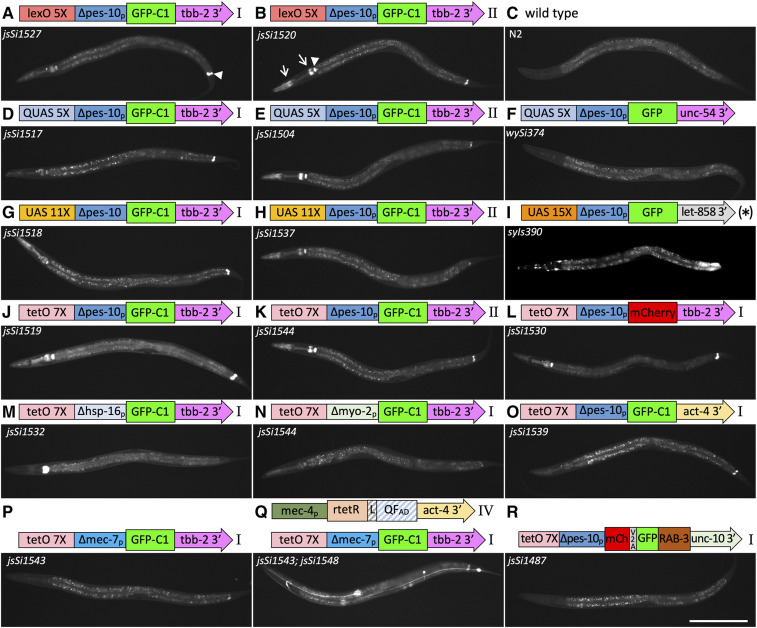
Background observed in bipartite reporter constructs. Widefield epi-fluorescence images of L4 animals taken using a 10× air lens with accompanying schematic diagrams of reporter constructs. (A–K) Animals homozygous for RMCE insertions of four distinct reporter constructs at landing site I and II as well as a wild-type control, a QUAS integrant isolated using MosSCI (Wei et al. 2012), and a multi-copy (*) UAS 15× integrated array (Wang et al. 2017). Background signal in the absence of a driver is observed in the rectal gland cells rect_VL, rect _VR and rect_D in the tail (e.g., arrowhead in A), the pharyngeal/intestinal valve cells (e.g., arrowhead in B), and portions of the pharynx (e.g., arrows in B). Signal in the intestine is background autofluorescence (except for the posterior intestinal signal in (I). (L–R) Background levels in RMCE insertions at landing site I in cases where the fluorescent protein, the 3′ UTR sequences or the basal promoter of the tet0 7X ∆pes-10 GFP-C1 tbb-2 3′ reporter construct have been replaced. (P and Q) Comparison of tetO reporter background levels in presence and absence of a mec-4 driver showing that background in not amplified by the presence of doxycycline. Diagrams of constructs are not to scale. See Figures S5 and S6 for detailed diagrams. All images are taken under identical conditions (500 msec exposure). Detailed head and tail images are presented in Figures S9 and S10. Bar, 200 µm.

