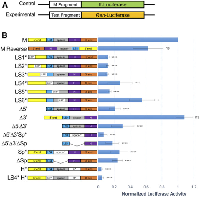
Figure 4.
Promoter analysis of the M fragment in transfected S2 cells. (A) Schematic of the control (M-firefly) and experimental (Test-Renilla) luciferase constructs. (B) Left: Schematics of the M fragment and derivatives used as test sequences in the promoter assay. See Figure 2A for details. Right: Luciferase activities of the indicated test sequences, normalized to the M-firefly luciferase activity. The M fragment has promoter activity in both orientations. Deletion of the 3′ end does not affect promoter activity. All other deletions or mutations affect promoter activity, with mutating the BEAF binding site having the strongest effect (H*: ∼50-fold) and LS6* having the weakest effect (∼2-fold). Three biological replicates were done, with SD indicated by the whiskers. One-way ANOVA P-values are indicated by asterisks (*P < 0.05; ****P < 0.0001; ns: not significant).

