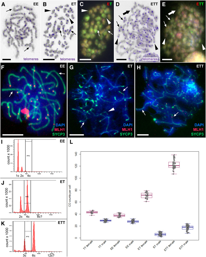
Figure 4.
Male meiotic spreads at metaphase I and pachytene stages. (A, B, and D) Giemsa-stained chromosomes (gray) with FISH-labeled telomeres (blue) in C. elongatoides, ET and ETT hybrid males, respectively. (C and E) The same metaphases as in (B and D) of hybrids after comparative genomic hybridization revealing the origins of individual chromosomes (red colour chromosomes correspond to C. elongatoides and green ones to C. taenia). Thin arrows indicate exemplary cases of bivalents, arrowheads exemplary univalents, and thick arrows exemplary cases of multivalents. (F–H) Meiotic spreads at pachytene stage of C. elongatoides (F), diploid ET hybrid (G), and triploid ETT hybrid (H) males stained with DAPI (blue). Synaptonemal complexes were immunolabeled with antibodies against SYCP3 protein (green) and MLH1 protein (red). Arrows indicate exemplary bivalents, arrowheads show examples of abnormal pairing and failures of bivalent formation. Bar, 10 mm. (I–K) Flow cytometry results of testes of C. elongatoides (I), diploid ET hybrid males (J), and triploid ETT hybrid males (K). (L) Diagram shows the average frequencies of crossovers (COs) per cell in studied genomotypes (males and females indicated in blue and red, respectively).

