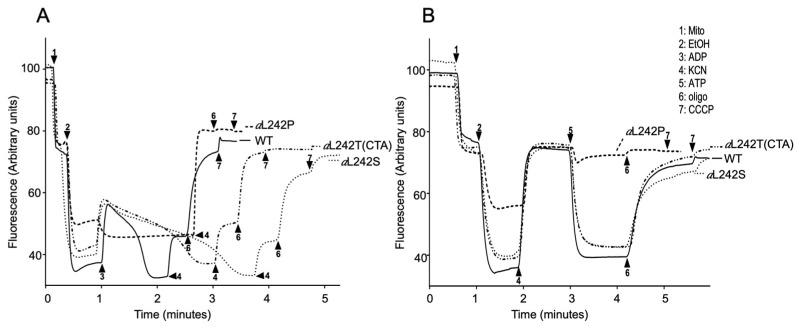
Figure 4.
Mitochondrial membrane potential. Variations in mitochondrial ΔΨ were monitored by fluorescence quenching of Rhodamine 123, using intact, osmotically-protected, mitochondria from wild type (WT) and mutant strains aL242P, aL242T and aL242S grown for 24 h at 28 °C in rich galactose liquid media until a density of 2-3 OD600nm (these mitochondrial preparations are the same as those used in Figure 2). The tracings in (A) show how the mitochondria respond to externally added ADP, those in (B) reflect ATP-driven proton-pumping by ATP synthase. The additions were 25 μg/mL Rhodamine 123, 0.15 mg/mL Mito, 10 μL EtOH, 75 μM ADP, 2 mM KCN, 0.2 mM ATP, 4 μg/mL oligo, and 4 μM CCCP. The shown fluorescence tracings are representative of at least three experiments.

